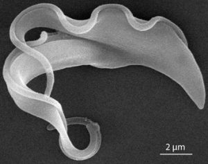
In the Laboratory of Cell Motility we study the cytoskeleton of the eukaryotic cell. We are particularly fascinated by the evolutionarily conserved microtubule-based organelles, the cilia and the flagella (both terms are interchangeable). Their beating drives motility of sperms and many parasitic and free living unicellular organisms. Moreover, in our body ciliary beating is crucial for mucociliary clearance and cerebrospinal fluid flow. Non-motile cilia, the so-called primary cilia, are present on almost every human cell, where they have critical signaling and sensory roles (Fig. 1). Thus, malfunctions of flagella and cilia lead to multi-symptomatic disorders collectively referred to as ciliopathies.
In our laboratory we attempt to answer some of the basic questions of the flagellum/cilium biology. We are intrigued by the complexity of these organelles, which consist of over a thousand protein species organized in a highly defined manner. How does the cell form such a complex yet highly organized organelle? Is there a protein turnover in these long, chemically and mechanically stable structures? How do individual constituents cooperate to achieve the highly coordinated behaviour giving rise to the flagellar beat?
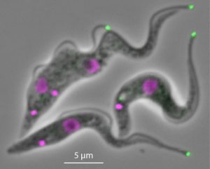
To address these questions we employ molecular biology, cell biology and biochemistry approaches to study ciliated mammalian cells and tissues, having the potential to provide insights into causes of ciliopathies. The mammalian experiments are inspired by our work on the protozoan Trypanosoma brucei. This important human parasite possesses a flagellum which is critical for cell morphogenesis, in addition to cell motility (Hayes & Varga et al., J. Cell Biol., 2014) (Fig. 2).
Crucially, T. brucei is the most experimentally tractable flagellated/ciliated organism. As such it allows for rapid development of technology and identification of proteins critical for particular flagellar processes. Studying two phylogenetically distant models- trypanosomes and mammals- also facilitates understanding of the evolution of these fascinating organelles.
The major project ongoing in our laboratory focuses on the distal end of the flagellum, the sole place of the flagellum cytoskeleton assembly. We develop biochemical approaches to identify proteins specifically localizing to this region of the flagellum (Varga et al., PNAS 2017) (Fig. 3). This allows us to study in vivo how do these proteins and structures they constitute orchestrate processes of flagellum cytoskeleton assembly and maintenance.
Moreover, we employ in vitro reconstitution assays using purified cytoskeletal proteins, akin to those described in Varga & Leduc et al., 2009, Cell, enabling us to gain a deep mechanistic insight into biochemical activities of the studied flagellar proteins.


